

By far the most common tumour you will see is osteosarcoma, in both dogs and cats, although it’s far more rare to see a cat with a bone tumour. However, if you see bone lesions, bear in mind that it might not be OSA, it could be another type of primary bone tumour, something benign, or even a fungal disease! This is why it’s important to actually get a diagnosis for these patients! We look at the appearance of the lesion as well as location, to give us clues as to what it might be, but ultimately need to sample it to be sure!
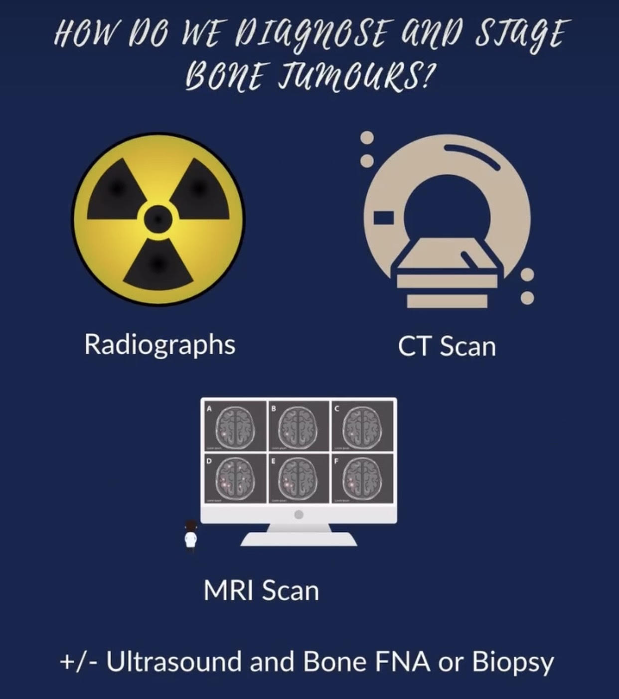
Primary bone tumours tend to be incredibly painful, so please consider analgesia if needed, even before we get our results of tests back. We may wish to perform radiographs or CT scanning of the thorax, CT scans are going to pick up nodules when they’re 2-3mm in comparison to 7-8 with radiographs, so it is generally more accurate. We may wish to do abdominal ultrasound, it’s rare that these metastasise to organs, but sometimes we will CT the abdomen and find concurrent issues that need investigation. We prefer to have a sample sent for cytology or histopathology, whether that’s fna or biopsy of a bone lesion, but take care as we don’t want to cause a pathologic fracture either!
 MOST COMMON BONE TUMOURS in dogs
 MOST COMMON BONE TUMOURS in dogs
Osteosarcoma
Chondrosarcoma
Haemangiosarcoma
Fibrosarcoma
Multilobular Osteochondrosarcoma (MLO)
Metastasis to bone from other tumours
MOST COMMON BONE TUMOURS in cats
Osteosarcoma
Fibrosarcoma
Chondrosarcoma
Haemangiosarcoma (rare)
Metastasis to bone from other tumours

OTHER/BENIGN/DIFFERENTIALS
Osteoma
Multiple cartilaginous exostosis (MCE)
Bone cysts
To consider with bone changes:
Fungal disease
Bacterial infection
CLINICAL SIGNS – BONE CANCER
- Lameness
- Pain (benign bone tumours such as osteomas generally non-painful)
- Swelling at primary site
- History of mild trauma and worsening lameness
- Rare – acute severe lameness due to pathologic fracture
- Axial bone lesions dependant on area-swelling, dysphagia or pain (oral), exopthalmos, facial deformity, neitological signs (spinal)

HOW DO WE DIGGNOSE AND STAGE
BONE JUmBURS?
Radiographs
CT Scan
MRI Scan
+/- Ultrasound and Bone FNA or Biopsy
REFERENCES:
Kudnig, S. T., & Séguin, B. (2012).
Veterinary Surgical Oncology. John Wiley & Sons, Ltd.
Withrow, S. J., Vail, D. M., Thamm,
D. H., & Liptak, J. M. (Eds.). (2019).
Withrow and MacEwen’s Small
Animal Clinical Oncology (6th ed.).
Elsevier.